Where Do Lumbar Splanchnic Nerves Synapse
The spinal accessory nerve is responsible for controlling the muscles of the neck along with cervical spinal nerves. Sympathetic chain in sacral region.

Lumbar Splanchnic Nerves An Overview Sciencedirect Topics
Here the fibers leave the spinal nerves enter the presacral area and branch out into three main locations.
. What do lumbar splanchnic nerves typically innervate. Some of its fibers do not synapse here but pass directly to the medulla of the adrenal gland which they innervate. B consist of axons that synapse in collateral ganglia.
The preganglionic cell bodies that contribute to parasympathetic pelvic splanchnic nerves originate from the lateral horn lateral. Provide sympathetics to handgun and pelvic viscera. D originate from first-order neurons located in the upper five thoracic segments of the spinal cord.
Anatomical terms of neuroanatomy. It should be noted that splanchnic nerves carry sympathetic fibers except for pelvic splanchnics which are parasympathetic. With what do post-ganglionic fibers run to target viscera.
The corresponding postsynaptic fibers then modulate the. A form part of the rami communicantes. From these prevertebral ganglia the postganglionic fibres supply organs in the pelvis lower abdomen and lower limb.
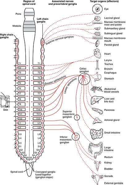
The lumbar splanchnic nerves arise from the upper lumbar levels and terminate in the inferior mesenteric and hypogastric ganglia. What do lumbar splanchnic nerves supply. 7 Splanchnic nerves A connect chain ganglia.
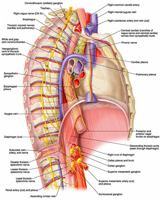
The lumbar splanchnic nerves travel through the lumbar sympathetic ganglion but do not synapse there. What prevertebral ganglia do least splanchnic nerves synapse on. Its fibers synapse with their postganglionic counterparts in the celiac ganglia or in the aorticorenal ganglion.
The large intestine and the kidneys are the main targets of this nerve with the pelvic plexus getting contributions when the nerve terminates in the inferior mesenteric and hypogastric ganglia. The nerve travels inferiorly lateral to the greater splanchnic nerve. E control abdominopelvic viscera.
It courses lateral and in parallel to the greater splanchnic nerves over the anterior surface of the spine and enters the abdomen in the same manner - by traversing the ipsilateral diaphragmatic crus. Lumbar Splanchnic Nerve. It synapses in the aorticorenal ganglion.
See Appendix 2-6 and see color plates. The nerve modulates the activity of the enteric nervous system of. Splanchnic nerves myelinated are those which come from the spinal cord pass through the sympathetic chain they do NOT synapse there like the rami communicans and proceed to synapse elsewhere.
The site of synapse is found in the inferior mesenteric ganglion and the postsynaptic fibers innervate the smooth muscle and glands of the pelvic viscera and hindgut. Where do sacral splanchnic arise from. Where do lumbar splanchnic nerves synapse.
Its preganglionic sympathetic fibers run medially and downwards to join the aortic plexus where they synapse in the ganglia there and then the postganglionic fibers are distributed to the vessels smooth muscles and. The lumbar splanchnic nerves are splanchnic nerves that arise from the lumbar part of the sympathetic trunk and travel to an adjacent plexus near the aorta. These nerves contain preganglionic sympathetic and general visceral afferent fibers.
B innervate glands and smooth muscles in the body wall. This nerve originates from thoracic ganglion 12 and synapses within ganglia. Anatomical terms of neuroanatomy.
Contribute to preaortic abdominal plexuses celiac superior mesenteric intermesenteric superior hypogastric splanchnic pelvic. The first is the lower thoracic splanchnic nerves that contain greater lesser and least branches which originate from the thoracic division of the sympathetic trunks and the second one is the lumbar splanchnic nerves that arise from the lumbar division of the sympathetic. Sensory nerves sometimes called afferent nerves.
Referred pain is a phenomenon of feeling pain at a site other than the site of the painful stimulus origin. The lesser splanchnic nerve arises from thoracic ganglia 10 and 11 Standring et al 2008. The anatomical arrangement of the roots of the cranial nerves observed from an inferior view.
This pelvic plexus also contains parasympathetic nerves. From these prevertebral ganglia the postganglionic fibres supply organs in the pelvis lower abdomen and lower limb. Pain Procedures in Clinical Practice Third Edition 2011.
C rejoin the spinal nerves. Where do lumbar splanchnic synapse. Lesser splanchnic nerve.
Ventral primary rami of spinal nerves S2-S4 cell bodies are located in the lateral horn gray of the sacral spinal cord. Depending on their function nerves are known as sensory motor or mixed. What do lumbar splanchnic nerves innervate.
Nerve nerv a macroscopic cordlike structure of the body comprising a collection of nerve fibers that convey impulses between a part of the central nervous system and some other body region. The lumbar splanchnic nerve one on each side of the body arises from the upper two ganglia of the lumbar part of the sympathetic chain L1L2. Figure 1332 The Cranial Nerves.
The lumbar splanchnic nerves arise from the upper lumbar levels and terminate in the inferior mesenteric and hypogastric ganglia. The pelvic splanchnic are preganglionic nerves that arise from the spinal cord lateral horn of sacral segments of the S2 S3 and S4 nerve roots though the greatest contribution of these fibers is usually from the S3 nerve. Instead they synapse at the inferior mesenteric ganglion and innervate the smooth muscle lining the large intestines kidney bladder glands of the hindgut and pelvic viscera.
There are four of these on each side. Least splanchnic nerve. What kind of fibers do most sacral splanchnic carry.
The lumbar splanchnic nerve supplies sympathetic innervation to the glands and smooth muscles of the hindgut and pelvic viscera. The first is the lower thoracic splanchnic nerves that contain greater lesser and least branches which originate from the thoracic division of the sympathetic trunks and the second one is the lumbar splanchnic nerves that arise from the. Despite numerous studies based on.
Are splanchnic nerves Postganglionic. The abdominopelvic splanchnic nerves which form the sympathetic aspect consists of three branches. Where do the greater lesser and least splanchnic nerves originate.
C are formed of parasympathetic fibers. Handgut and pelvic viscera. Structure and Function.
The three splanchnic nerves synapse at the celiac ganglia which lie anterior and anterolateral to the aorta. Where do the splanchnic nerves synapse. They originate from L1 and L2.
The hypoglossal nerve is responsible for controlling the muscles of the lower throat and tongue. The lesser splanchnic nerves terminate by synapsing within the aorticorenal or superior mesenteric ganglia. E control sympathetic function of structures in the thorax.
The majority of the splanchnic fibers join the inferior hypogastric plexus while the smaller portion of the fibers joins the hypogastric nerves and travels with them to the superior hypogastric plexus. The splanchnic nerves are paired visceral nerves nerves. These nerves contain preganglionic sympathetic and general visceral afferent fibers.
Sacral splanchnic nerves are parasympathetic or. It arises from a pathological mixing of nociceptive processing pathways for visceral and somatic inputs.

Splanchnic Nerves An Overview Sciencedirect Topics

Clinical Anatomy Of The Splanchnic Nerves Haroun Moj Anatomy Physiology

Sympathetic Nervous System Page 2

Anatomy Back Splanchnic Nerve Article

Splanchnic Nerve Modulation In Heart Failure Mechanistic Overview Initial Clinical Experience And Safety Considerations Fudim 2021 European Journal Of Heart Failure Wiley Online Library

Splanchnic Nerves An Overview Sciencedirect Topics

Clinical Anatomy Of The Splanchnic Nerves Haroun Moj Anatomy Physiology
No comments for "Where Do Lumbar Splanchnic Nerves Synapse"
Post a Comment